Disease, diagnostics and the care of senior pets
by Sandra Wells, NationWide Laboratories
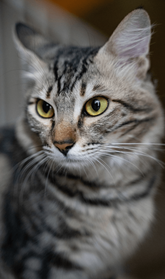
Case study: Possible feline injection-site sarcoma in a 13-year-old domestic shorthair cat
by Dante Meza Ruiz Associate Pathologist, NationWide Laboratories
Pathology Focus
Age is not a disease. Yet, as pets grow older, they become more susceptible to a range of health issues that can impact their quality of life. Thanks to advances in veterinary medicine, including improved surgical techniques, more advanced diagnostics, better nutrition and more, pets are living longer, healthier lives.
Over recent years, the lifespan of dogs has increased by approximately five percent while the longevity of cats has seen an even more significant rise, with purebred felines living about nine percent longer and mixed breeds nearly fourteen percent longer.1
Many veterinary practices offer nurse-led ‘senior’ clinics specifically designed to address the unique needs of these older pets, but when is a pet considered senior? With significant variations in breed, type and size, there is no universally fixed age. However, the American Animal Hospitals Association (AAHA) defines the senior stage as the last 25 percent of the
expected lifespan,2 meaning it occurs earlier for large and giant breeds than for smaller ones.
Ageing is a gradual and irreversible pathophysiological process. At the tissue level, this process manifests as pathological changes such as atrophy, fibrosis and fatty infiltration, all of which contribute to the decline in tissue, cellular and organ function.3 While the ageing process itself is not a disease, these changes inevitably increase the risk of age-related health conditions.
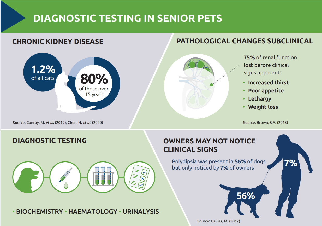
Importantly, these pathological processes often develop silently, with many age-related diseases remaining subclinical for extended periods. For instance, chronic kidney disease (CKD) is a relatively common condition, especially in older cats, but it may not become clinically apparent until approx. 75 percent of renal function is lost.4
Even when signs of age-related disease are present, owners may not always recognise or report them. A study involving 45 dogs over nine years of age attending a first-opinion practice, revealed that owners frequently overlooked or misunderstood important clinical signs. For example, only seven percent of owners reported increased thirst, despite it being present in 56 percent of the dogs studied.5
Additionally, none of the owners noticed weight loss in their pets, even though all dogs with a low body condition score, and some with ideal scores, had lost weight prior to screening. This discrepancy highlights that, alongside clinical examination, routine monitoring through laboratory diagnostics has an important role to play.
Earlier identification of health issues in senior pets can extend both the quality and quantity of life. With this in mind, the primary aim of diagnostic screening is to:
Detect subclinical disease
Enable early investigation, diagnosis and intervention
Identify risk factors
Identify problems that require ongoing monitoring
If no abnormalities are detected, the screening results can serve as a valuable baseline for future comparison, enabling more precise monitoring of changes over time.
While the benefits of a thorough clinical examination cannot be overstated, laboratory diagnostics offer key insights that are not always apparent from a physical examination alone. A typical geriatric profile includes parameters to assess renal function, thyroid function, glucose metabolism and other key health indicators. In the aforementioned study of 45 dogs, an average of seven to eight health issues were identified per dog and previously unrecognised problems were found in 80% of cases.
Chronic kidney disease (CKD) is one of the most common diagnoses in older cats, with its prevalence rising significantly with age. In fact, studies have shown that while CKD affects approximately one percent of the general cat population,6 its prevalence jumps to as high as 80 percent in those over 15 years.7
Traditional markers like serum creatinine are commonly used to assess renal function, but they have limitations, particularly in the early stages of CKD. In line with the clinical picture, creatinine levels may remain within normal ranges until approximately 75 percent of renal function is lost, making early detection challenging.
Originally published in the Veterinary Edge, October 2024
This is where symmetric dimethylarginine (SDMA) proves invaluable. SDMA is a more sensitive biomarker for renal function that can detect decreases in glomerular filtration rate (GFR) earlier than creatinine; SDMA levels increase when GFR decreases by approximately 40 percent, providing a more timely indication of renal dysfunction. Another advantage of SDMA is that it is less impacted by loss of lean body mass than creatinine, making it a potentially more reliable marker in older pets.
The International Renal Interest Society (IRIS) recognises the importance of SDMA in the early detection and staging of CKD. According to the IRIS guidelines,8 persistently elevated SDMA levels, even when serum creatinine is within normal limits, may indicate early-stage CKD (IRIS stage 1).
In reality, creatinine and SDMA levels should be assessed in tandem when investigating or screening for renal disease. In addition, levels of both parameters should be assessed on at least two occasions in a stable, hydrated patient to confirm persistent elevation.
As pets live longer, the need for proactive healthcare becomes increasingly important. Ageing, while not a disease in itself, inevitably leads to physiological changes that heighten the risk of various clinical conditions. Diagnostic testing plays a pivotal role in identifying these issues early, allowing for timely interventions that can significantly enhance both the quality and quantity of life in senior pets.
Click here for references
To find out more about NationWide Laboratories and all the services they offer, visit - https://nwlabs.co.uk or click on the video above.
Pathology Case Study
By Dante Meza Ruiz Associate Pathologistat NationWide Laboratories,DVM, M.Ed., DVSc, Dipl. ACVP/ASVCP
A 13-year-old spayed female domestic shorthair cat was presented to a veterinary clinic with a history of a large mass, approximately 8 – 10 cm in size, with an irregular texture. The mass was located on the left caudal border of the scapula and exhibited firm attachment, extending approximately 2 cm across the spine. A fine-needle aspiration (FNA) was performed, and the sample was submitted to NationWide Laboratories for evaluation.
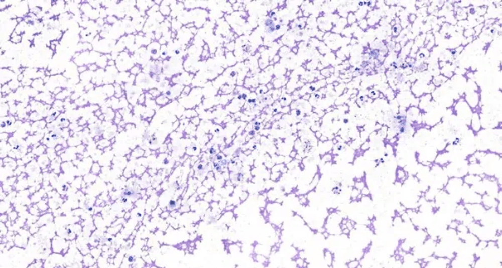
The cytologic evaluation revealed a poorly cellular and mildly hemorrhagic sample, characterised by low numbers of poorly preserved, often lysed neutrophils, a few vacuolated macrophages, and rare small lymphocytes and plasma cells within a dense eosinophilic proteinaceous background (Figure 1).
Findings were consistent with the aspiration of proteinaceous fluid and mild mixed inflammation, predominantly neutrophilic, potentially associated with a cyst or cystic neoplasm. However, the exact identity of the lesion could not be determined from the sample.
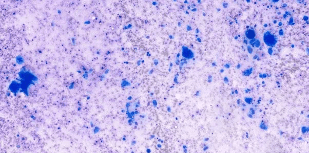
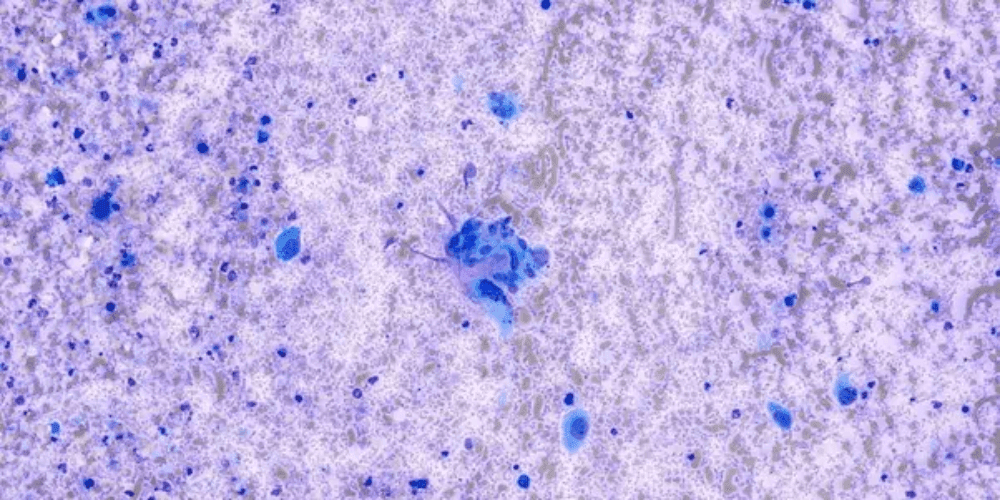
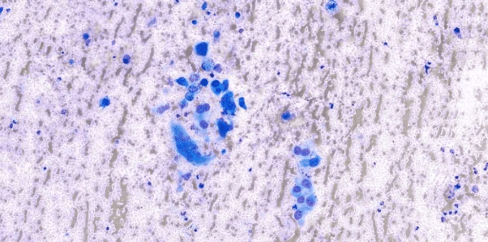
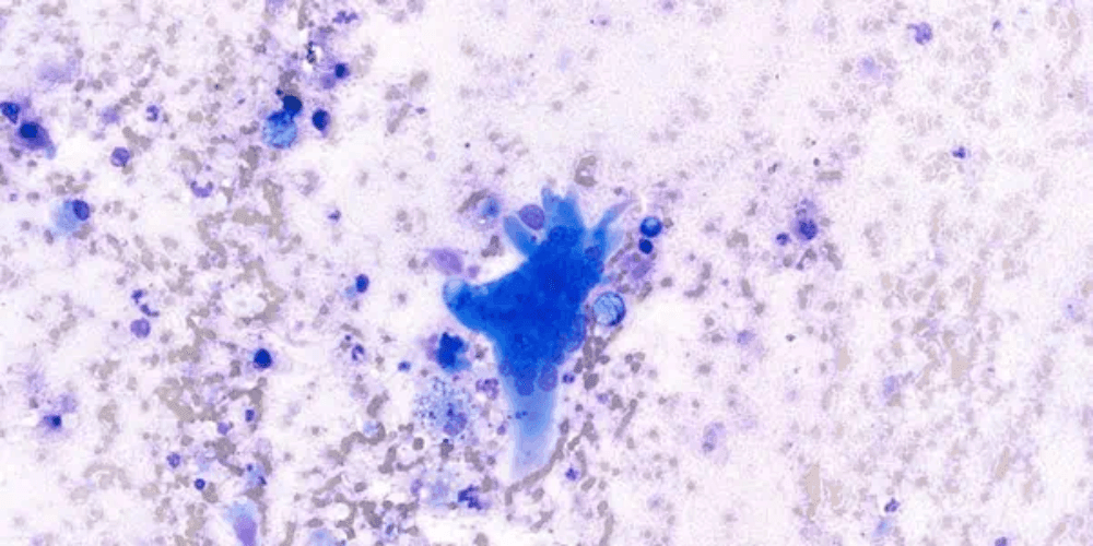
A few days later, a second FNA was performed, and cytologic evaluation revealed a densely eosinophilic, proteinaceous background containing numerous neutrophils and highly vacuolated macrophages. Additionally, several spindle-shaped cells were observed, arranged individually, in loose aggregates, and occasionally associated with pink extracellular material. These spindle-shaped cells exhibited moderate to large amounts of wispy grey-blue cytoplasm with variably distinct borders, oval nuclei with finely stippled chromatin, and one to multiple prominent round nucleoli. Marked anisocytosis and anisokaryosis were noted, along with frequent giant multinucleated cells, some containing more than 20 nuclei. Occasional karyomegaly, anisonucleosis, and bizarre mitotic figures were also identified (Figures 2-5: Photomicrograph of direct smears from a fine-needle aspirate of a mass in a cat, stained with Leishman stain).
A diagnosis of sarcoma with mixed inflammation, predominantly neutrophilic, was established. Considering the location, pronounced cellular pleomorphism, marked dysplastic features, and the presence of multinucleated giant cells, the primary differentials include injection-site sarcoma and undifferentiated pleomorphic sarcoma. However, the possibility of a reactive process or other mesenchymal tumours was not excluded.
Soft tissue sarcomas (STS) are a frequently diagnosed group of malignant tumours in domestic cats, encompassing various histological subtypes and exhibiting diverse biological behaviours. The terminology surrounding these tumours can be challenging, due in part to the extensive range of terms used in the literature. Additionally, the existence of feline injection-site sarcomas (FISS) further complicates the classification and understanding of these neoplasms in cats1.
FISS are among the most serious vaccine-associated adverse events. These tumours typically develop at sites commonly used for vaccinations and injections, including the interscapular region, the lateral thoracic or abdominal wall, the lumbar region, and the area around the semimembranosus and semitendinosus muscles. The exact pathogenesis of these sarcomas remains unclear, and there is no definitive causal relationship or direct link with vaccination. However, chronic inflammatory reactions at the injection site are believed to serve as a trigger for subsequent malignant transformation2.
Most feline injection-site sarcomas (FISS) are fibrosarcomas, although other malignancies, including osteosarcomas, chondrosarcomas, rhabdomyosarcomas, malignant fibrous histiocytomas, and myofibroblastic sarcomas, have been rarely reported. FISS are generally more aggressive than sarcomas at other sites. The metastasis rate is relatively low, with the lungs being the most common site of metastasis, followed by regional lymph nodes and abdominal organs3.
The prognosis of FISS is primarily influenced by tumour size, with radical excision being crucial for a favourable outcome. Prognosis is also affected by the tumour’s location, its accessibility to surgery, and the ability to achieve tumour-free margins. Therefore, pre-operative diagnostic imaging is essential to assess the full extent of the tumour. Prognosis improves when radical surgery is combined with additional therapeutic options, such as radiotherapy or immunotherapy3.
Read more case studies from NationWide Laboratories
References