Pathology case studies:
Lymphangiosarcoma in a CatBy Karina Fresneda, Anatomic Pathologist at NationWide Laboratories, DVM DiplACVP
Digital adenocarcinoma in a domestic shorthair catBy Sofia Clara Sacco, Anatomic Pathologist at NationWide Laboratories, DVM, PhD
Pathology Case Study
By Karina Fresneda, Anatomic Pathologist at NationWide Laboratories, DVM DiplACVP
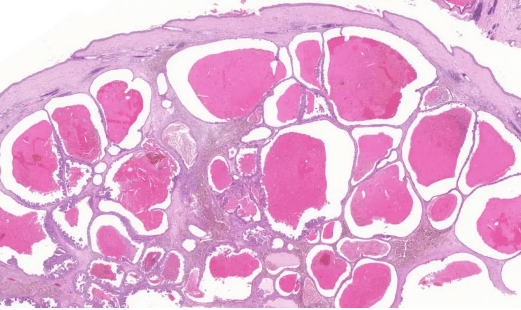
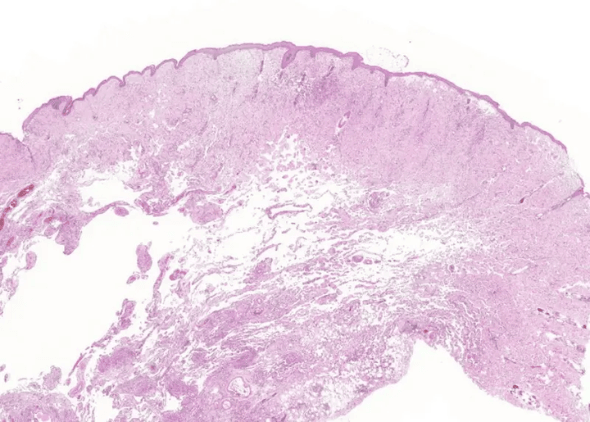
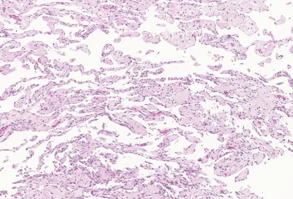
We received a skin and subcutaneous tissue sample from the caudoventral abdomen of an 8-year-old domestic short hair cat.
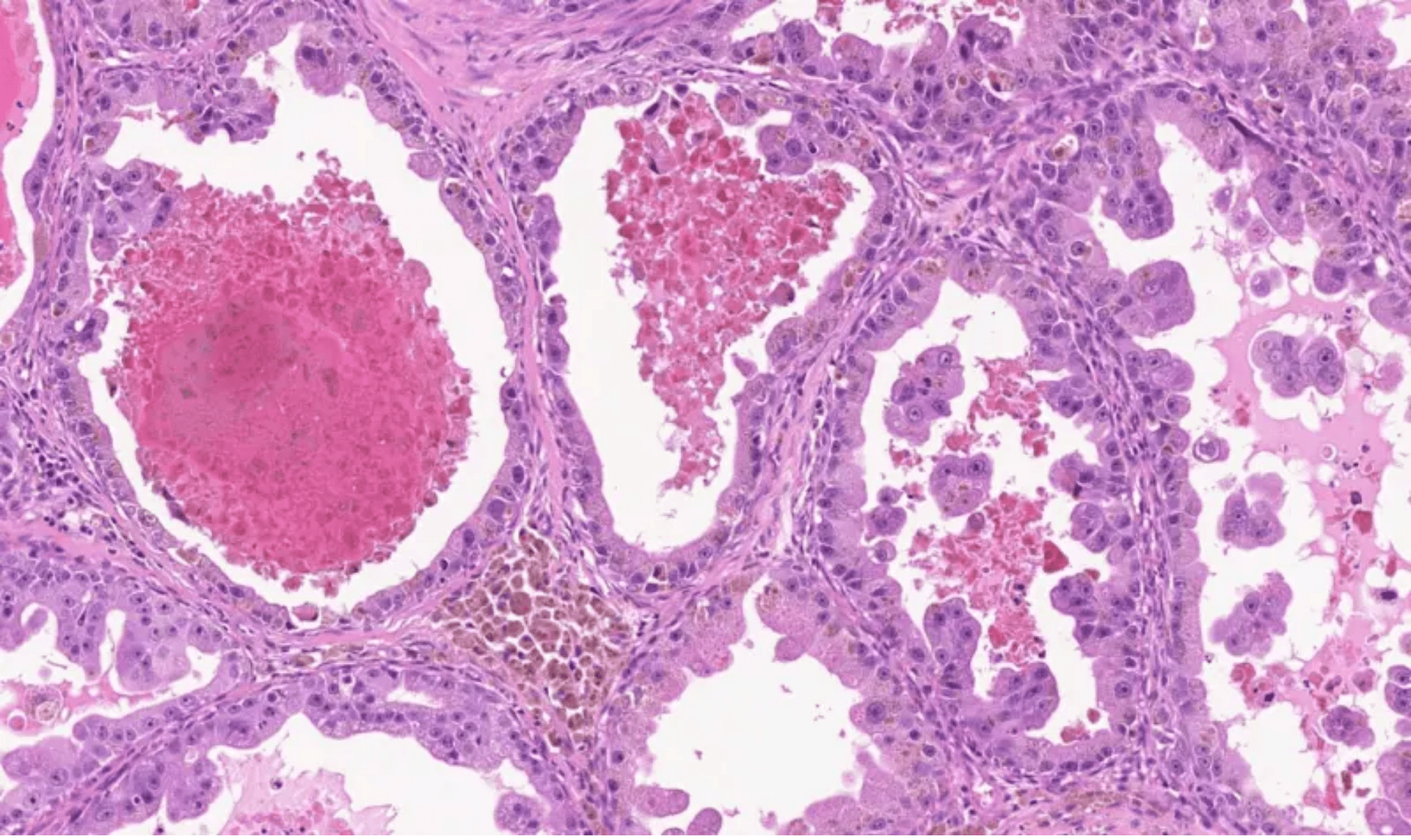
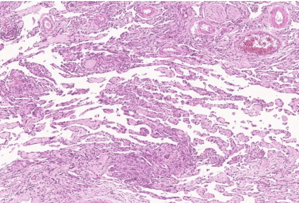
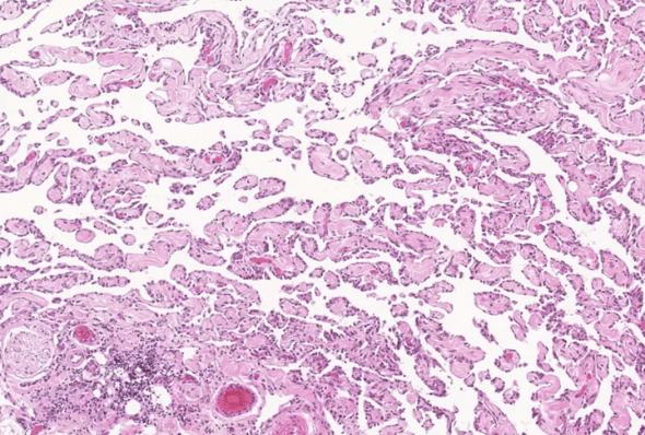
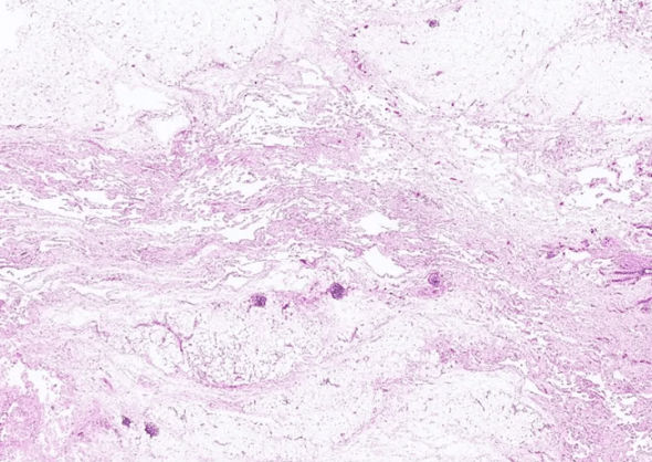
Microscopic examination revealed a non-demarcated, unencapsulated, and infiltrative proliferation of irregular vascular channels. These channels contained few to no erythrocytes and dissected between dermal collagen bundles, adipose tissue, and muscle fibers. They were lined by a single layer of round to oval endothelial cells, which occasionally protruded into the vessel lumina.
The neoplastic cells exhibited basophilic cytoplasm and round to elongated nuclei, occasionally with more than one nucleolus. Mitotic figures were rare, with 0–1 per 10 high-power fields (HPFs). These histopathological features were consistent with a diagnosis of well-differentiated lymphangiosarcoma.
Lymphangiosarcoma is a rare malignant vascular neoplasm in cats, with no known breed predisposition. It typically arises within the dermis and subcutis and may occur at any anatomical site. However, it is most frequently reported in the subcutaneous tissues of the ventral thorax and abdomen, particularly the caudoventral abdominal wall. Interestingly, development of lymphangiosarcoma has also been documented in the ventrothoracic region following forelimb amputation in two feline cases.
Clinically, affected areas may present with poorly circumscribed, and highly infiltrative, extensive bruising and oedema. Draining tracts that exude haemorrhagic to serosanguineous fluid are often observed and may be associated with focal, erythematous, and hyperpigmented macules. In some cases, chylous pleural and peritoneal effusions have been reported, particularly involving the cranial mediastinum and mesentery, respectively. Regional lymph node involvement may occur.
Cytology is generally not diagnostic for this tumour, and definitive diagnosis relies on biopsy and histopathological evaluation. Histologically, lymphangiosarcomas are characterised by a poorly defined proliferation of spindle-shaped endothelial cells forming vascular clefts or cavernous spaces.
A key diagnostic feature distinguishing lymphangiosarcoma from hemangiosarcoma is the relative absence of erythrocytes within the abnormal vascular channels. Cytological atypia, abnormal vascular architecture, and infiltrative growth distinguish this neoplasm from normal vascular endothelial tissue. To avoid diagnostic ambiguity, the term angiosarcoma is sometimes used more broadly.
Immunohistochemistry can aid in differentiation: markers such as factor VIII and CD31 confirm endothelial origin, while PROX-1 supports lymphatic lineage.
Although metastasis is considered uncommon, the prognosis is guarded due to the tumour’s locally aggressive and recurrent nature. Currently, repeated surgical excision remains the only recognised treatment option.
Click here for references
Read more case studies from NationWide Laboratories
By Sofia Clara Sacco, Anatomic Pathologist at NationWide Laboratories, DVM, PhD
An 11-year-old neutered male Domestic Shorthair cat was presented to a veterinary clinic with a two-month history of a digital lump. Amputation of the affected digit was performed, and the sample was submitted to NationWide Laboratories for evaluation.
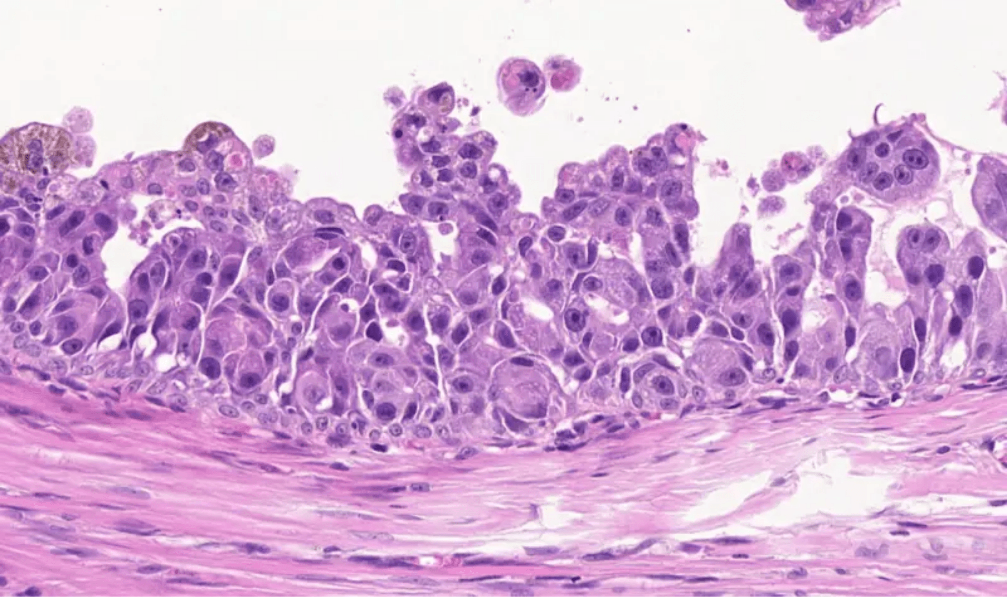
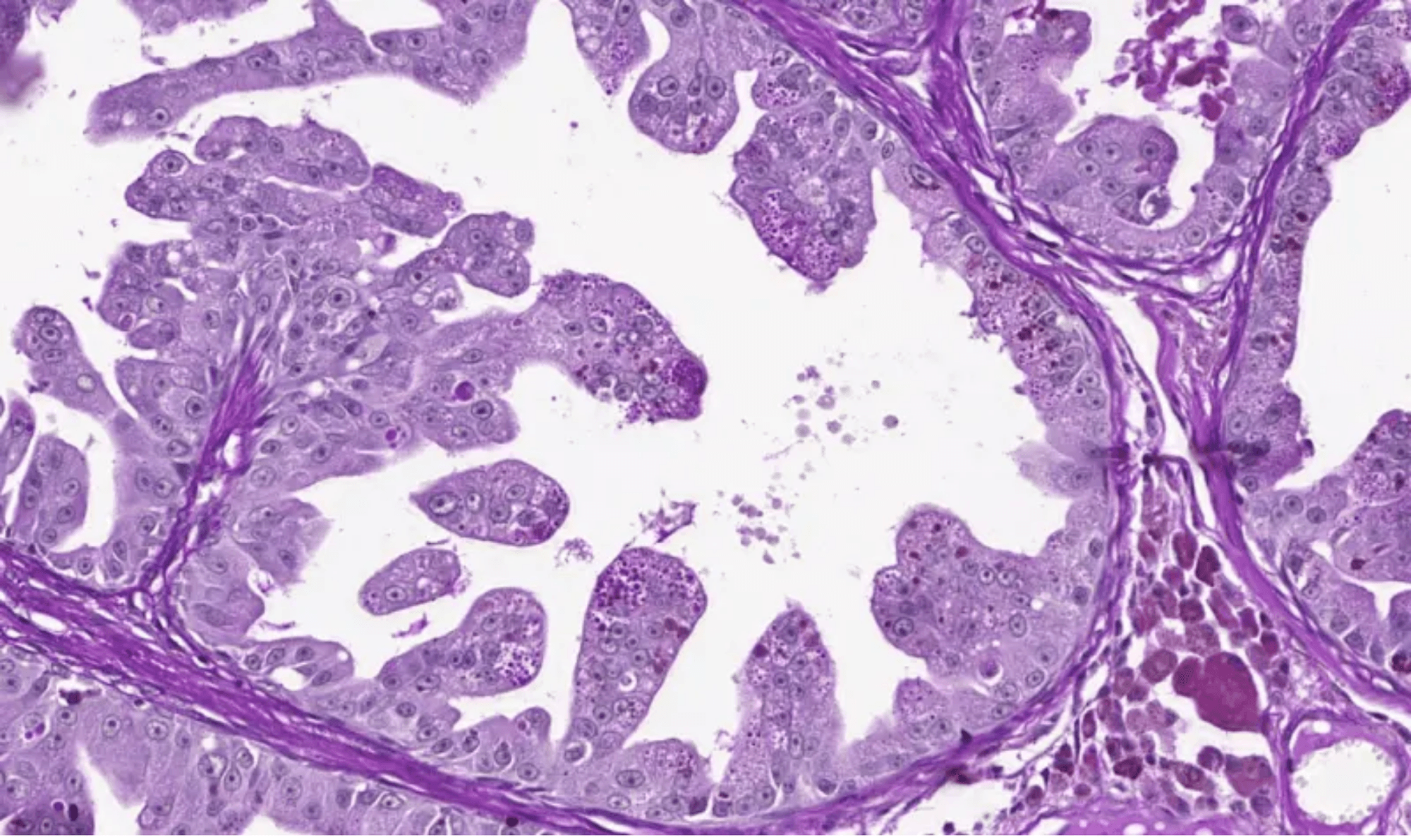
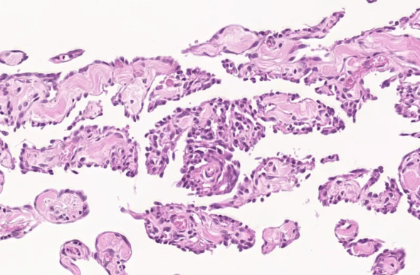
In the dermis, there was an unencapsulate moderately demarcated and invasive neoplasm composed of cells arranged in variably sized, irregular tubules and papillae, supported by a moderate amount of fibrous connective tissue (desmoplasia).
These tubules were lined by one to multiple layers of cuboidal to columnar neoplastic epithelial cells and contained central lakes of eosinophilic secretory material. The neoplastic cells showed moderate to abundant eosinophilic cytoplasm, oval nuclei, stippled chromatin and a single prominent magenta nucleolus. Moderate to marked anisocytosis and anisokaryosis were present. Six mitotic figures were observed per 10 HPF.
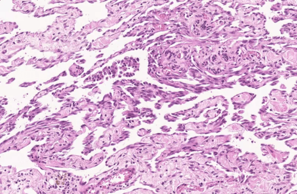
Interspersed within the neoplasm, there was a moderate inflammatory infiltrate composed predominantly of foamy macrophages and neutrophils, with fewer lymphocytes and plasma cells, and rare mast cells. The neoplastic cells were positive for periodic acid–Schiff (PAS) staining, both for intracytoplasmic material and for secretory material within the lumina.
A diagnosis of adenocarcinoma was established and based on the histological characteristics and PAS positivity of the cells; a metastatic neoplastic process was suspected.
This neoplasm was consistent with an adenocarcinoma, characterised by PAS-positive intracytoplasmic material and PAS-positive secretory material, raising the suspicion of a metastatic pulmonary carcinoma. The main differential diagnosis in this case is primary apocrine or eccrine carcinoma of the digit. Nevertheless, primary adenocarcinomas of the digit are typically PAS-negative. Immunohistochemistry is recommended as an additional diagnostic tool to help confirm the origin of the neoplasm.
Cutaneous metastasis of internal tumours is rare in cats. Pulmonary adenocarcinoma associated with feline lung–digit syndrome is the most common cause of tumour metastasis to the skin in this species; other possible primary sites include the mammary glands and gastrointestinal tract.
Feline lung–digit syndrome describes an unusual metastatic pattern observed with various types of primary pulmonary tumours, where metastases occur in the distal phalanges of the limbs. Primary lung tumours in cats are often not detected based on clinical signs related to the thorax; rather, many cases present with signs referable to distant metastases. In cats presenting with lameness or digital inflammation, lung–digit syndrome should be included in the list of differential diagnoses, even in the absence of respiratory signs. Primary pulmonary carcinoma should be considered in any middle-aged to elderly cat presenting with digital disease.